Upper Back Anatomy - Muscles Of The Upper Back And Neck Anatomy. Other important bones in the shoulder include: The upper back is the region below the cervical spine (neck) and above the low back (lumbar spine). The superficial and intermediate muscles do not develop in the back, and are classified as extrinsic muscles. It is like that for several reasons, all of which you can understand by looking at the anatomy of the thoracic spine. Skeletal muscle anatomy youtube 12 photos of the skeletal muscle anatomy youtube skeletal muscle anatomy youtube, human muscles, skeletal muscle anatomy youtube
Before giving our recommendations for upper back exercises, it's important to first go over the anatomy of the back musculature. Both the deltoid and the trapezius are firmly attached to the spine of the scapula. Related posts of anatomy of the back organs anatomy of human body organs. This muscle is located on the upper portion of the back anatomy, underneath the trapezius. The upper back muscles of the rhomboids and the trapezius are responsible for many of the movements of the scapula which in turn plays a huge role in the stability and mobility of the shoulder.
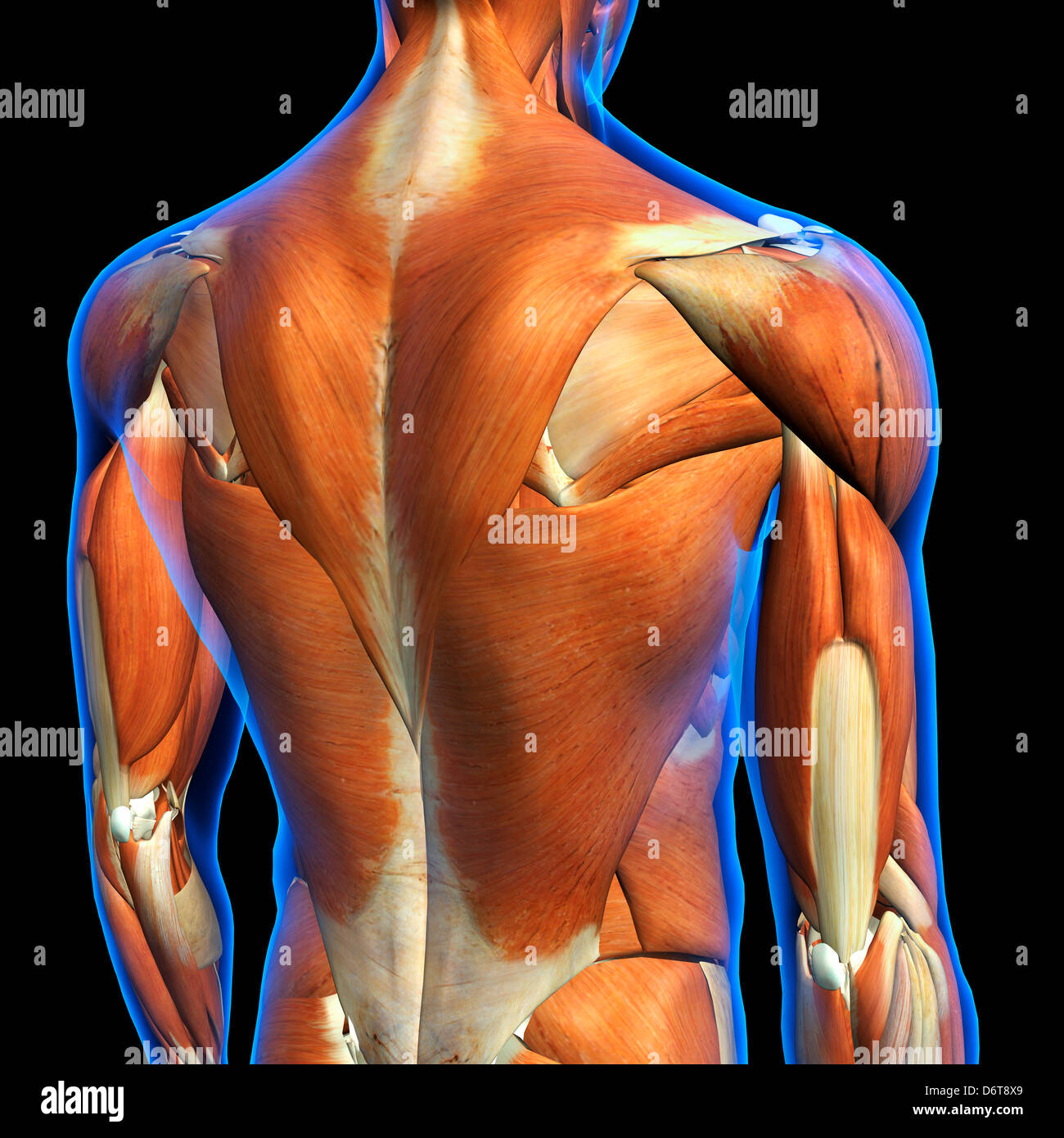
Male Upper Back Muscles Anatomy In Blue X Ray Outline Full Color 3d Computer Generated Illustration On Black Background Stock Photo Alamy from c8.alamy.com The lower back (lumbar vertebrae) allows for flexibility and movement in back bending (extension) and forward bending (flexion). The upper back is called the thoracic spine, and it is the most stable part of the spine. The cervical spine protects the nerves connecting to. It consists of seven vertebrae. It is very stiff, and the thoracic spine has a limited range of motion. The rib cage also anchors the bones of the head, neck, shoulders, and arms to the trunk of the body. Both the deltoid and the trapezius are firmly attached to the spine of the scapula. There is a set of muscles in the upper back (called the thoracic area) called the spinalis thoracis.
Anatomy of human body organs 12 photos of the anatomy of human body organs anatomy human body organ systems foldable, anatomy of human body and its functions, anatomy of human body drawing pdf, anatomy of human body parts, anatomy of human body quiz, human anatomy, anatomy human body organ systems foldable, anatomy.
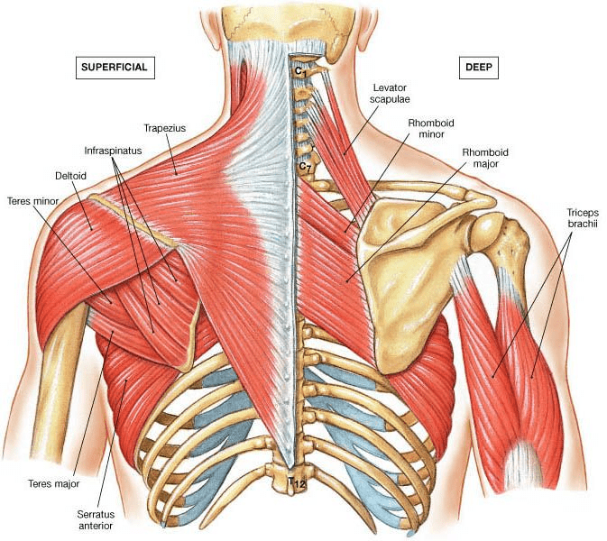
The lower back (lumbar vertebrae) allows for flexibility and movement in back bending (extension) and forward bending (flexion). The upper back has the most structural support, with the ribs attached firmly to each level of the thoracic spine and very limited movement. The bones of the chest and upper back combine to form the strong, protective rib cage around the vital thoracic organs such as the heart and lungs. The upper back is the region below the cervical spine (neck) and above the low back (lumbar spine). The rib cage also anchors the bones of the head, neck, shoulders, and arms to the trunk of the body. Powerful muscles that move the head and arms attach to these bones as well. It consists of seven vertebrae.it comprises the vertebral column (spine) and two compartments of back muscles; This is my video about the muscles of the back. The complexity of this region means that dysfunction can occur either due to injury or progressive pain and degeneration. The deltoid, teres major, teres minor, infraspinatus, supraspinatus (not shown) and subscapularis muscles (not shown) all extend from the scapula to the humerus and act on the shoulder joint. The main superficial muscles of the back are the following: It consists of seven vertebrae. The muscles of the back.
The lower back (lumbar vertebrae) allows for flexibility and movement in back bending (extension) and forward bending (flexion). This muscle is located on the upper portion of the back anatomy, underneath the trapezius. The complexity of this region means that dysfunction can occur either due to injury or progressive pain and degeneration. They originate from the vertebrae and insert into the scapulae. Skeletal muscle anatomy youtube 12 photos of the skeletal muscle anatomy youtube skeletal muscle anatomy youtube, human muscles, skeletal muscle anatomy youtube
Upper Back Pain Local Physio from www.local-physio.co.uk The cervical spine protects the nerves connecting to. The upper back is called the thoracic spine, and it is the most stable part of the spine. It runs from the neck to the upper back. The extrinsic (superficial) back muscles, which lie most superficially on the back. It does not permit twisting. They originate from the vertebrae and insert into the scapulae. The muscles of the chest and upper back occupy the thoracic region of the body inferior to the neck and superior to the abdominal region and include the muscles of the shoulders. The upper back muscles of the rhomboids and the trapezius are responsible for many of the movements of the scapula which in turn plays a huge role in the stability and mobility of the shoulder.
It is very stiff, and the thoracic spine has a limited range of motion.
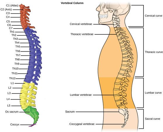
Connecting with the cervical spine above and the lumbar spine below, the thoracic spine runs from the base of the neck down to the abdomen. Related posts of upper back muscle diagram skeletal muscle anatomy youtube. The muscles of the back are a group of strong, paired muscles that lie on the posterior aspect of the trunk they provide movements of the spine, stability to the trunk, as well as the coordination between the movements of the limbs and the back muscles are divided into two large groups: It consists of seven vertebrae. This muscle is located on the upper portion of the back anatomy, underneath the trapezius. The trapezius and latissimus dorsi muscles connect the upper limb to the vertebral column. The complexity of this region means that dysfunction can occur either due to injury or progressive pain and degeneration. The superficial and intermediate muscles do not develop in the back, and are classified as extrinsic muscles. The cervical spine protects the nerves connecting to. The upper back is the region below the cervical spine (neck) and above the low back (lumbar spine). It is like that for several reasons, all of which you can understand by looking at the anatomy of the thoracic spine. The main superficial muscles of the back are the following: This is my video about the muscles of the back.
It runs from the neck to the upper back. Back anatomy the back is the body region between the neck and the gluteal regions. The bones of the chest and upper back combine to form the strong, protective rib cage around the vital thoracic organs such as the heart and lungs. The thoracic spine is the longest region of the spine, and by some measures it is also the most complex. It comprises the vertebral column (spine) and two compartments of back muscles;
Anatomy Of Upper Back Muscles Anatomy Drawing Diagram from physiologicnyc.com The cervical spine supports the weight and movement of your head and protects the nerves exiting your brain. The muscles of the chest and upper back occupy the thoracic region of the body inferior to the neck and superior to the abdominal region and include the muscles of the shoulders. The complexity of this region means that dysfunction can occur either due to injury or progressive pain and degeneration. The lumbar region of the spine, more commonly known as the lower back, is situated between the thoracic, or chest, region of the spine, and the sacrum. Anatomy of human body organs 12 photos of the anatomy of human body organs anatomy human body organ systems foldable, anatomy of human body and its functions, anatomy of human body drawing pdf, anatomy of human body parts, anatomy of human body quiz, human anatomy, anatomy human body organ systems foldable, anatomy. The main superficial muscles of the back are the following: These layers of back muscles help to mobilize and stabilize your trunk during your day to day activities. Skeletal muscle anatomy youtube 12 photos of the skeletal muscle anatomy youtube skeletal muscle anatomy youtube, human muscles, skeletal muscle anatomy youtube
Think of your spine as a tree trunk.
Extending from the base of the skull into the pelvis, it consists of 33 stacked bones known as vertebrae. They originate from the vertebrae and insert into the scapulae. The rhomboid muscle is activated as you bring and squeeze your scapula or shoulder blades back and together. The upper back has the most structural support, with the ribs attached firmly to each level of the thoracic spine and very limited movement. Upper back pain is most commonly caused by muscle irritation or tension, also called myofascial pain. Anatomy of back muscles your back consists of three distinct layers of muscles, namely the superficial layer, the intermediate layer, and the deep layer. The cervical spine protects the nerves connecting to. The muscles of the chest and upper back occupy the thoracic region of the body inferior to the neck and superior to the abdominal region and include the muscles of the shoulders. Related posts of anatomy of the back organs anatomy of human body organs. A pinched nerve in the upper back can cause, pain, numbness, and tingling in that area and other parts of the upper body, which can be uncomfortable. The muscles of the back. Related posts of upper back muscle diagram skeletal muscle anatomy youtube. Connecting with the cervical spine above and the lumbar spine below, the thoracic spine runs from the base of the neck down to the abdomen.